The microscope systems of the CBS Optical Imaging Facility are made freely available to members of CBS, MCB, and other departments. If you would like to become a registered user, please contact Steve Turney (sturney@mcb.harvard.edu or 6-9418). After you are registered, you are welcome to reserve time on the systems using SPINAL, the online scheduler.
Resources
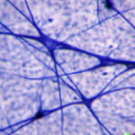
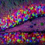
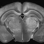
The imaging facility comprises three laser scanning confocal microscopes from Zeiss, two from Olympus, two TIRF microscopes from Nikon, a light-sheet microscope system from LiT, and a high-capacity slide scanner from Huron Digital Pathology. The microscope systems are fully motorized and allow automated acquisition of multi-channel images including Z-stacks, time series, and multi-position or overlapping tiles. The Olympus FV1000s and the Zeiss LSM 710 have upright stands. The remaining Zeiss and Nikon systems have inverted stands. The Nikon microscopes are equipped with incubation chambers, Perfect Focus Systems (automatic focus stabilization), and back-illuminated sCMOS cameras for fast and sensitive live cell time-lapse microscopy using TIRF, epifluorescence, or transmitted-light optics.
Documentation and Image Storage
A repository of user manuals and other microscope system documents and a web-based server for hosting and viewing whole slide images that are acquired with the slide scanner are available via the buttons to the left. Network storage is also available via FAS Research Computing (see FAQ below).
Microscope Systems


Summary of features and applications
| Tool | Slides | Live Imaging | 2 Photon | TIRF | Motorized Stage | CCD/sCMOS camera | TL Optics | Spectral Imaging |
|---|---|---|---|---|---|---|---|---|
| Huron LE-120 | included | not included | not included | not included | included | not included | included | not included |
| Nikon TE2000* | included | included | not included | included | included | included | included | not included |
| Nikon N-STORM* | included | included | not included | included | included | included | included | not included |
| Olympus FVA | included | not included | not included | not included | included | not included | included | included |
| Olympus FVB | included | included | included | not included | included | not included | included | not included |
| Zeiss 710 | included | included | not included | not included | included | not included | included | included |
| Zeiss 510 | included | included | not included | not included | included | included | included | not included |
| Zeiss Pascal | included | included | not included | not included | included | included | included | not included |
| LiTone XL | not included | included | not included | not included | included | included | not included | not included |
| Video-rate multiphoton | not included | included | included | not included | not included | not included | not included | not included |
Available laser lines on confocal / multiphoton systems
| Tool | 405nm | 440nm | 458nm | 488nm | 514nm | 543nm | 561nm | 594nm | 633nm | 2 Photon |
|---|---|---|---|---|---|---|---|---|---|---|
| Nikon N-STORM | included | not included | included | not included | not included | not included | included | not included | included | not included |
| Olympus FVA | included | not included | not included | included | not included | not included | included | not included | included | not included |
| Olympus FVB | included | not included | not included | included | not included | not included | included | not included | included | included |
| Zeiss 710 | included | included | included | included | included | not included | included | included | included | not included |
| Zeiss 510 | included | not included | included | included | included | included | not included | not included | included | not included |
| Zeiss Pascal | not included | included | included | included | included | included | not included | not included | not included | not included |
| Video-rate multiphoton | not included | not included | not included | not included | not included | not included | not included | not included | not included | included |
Imaging Rooms
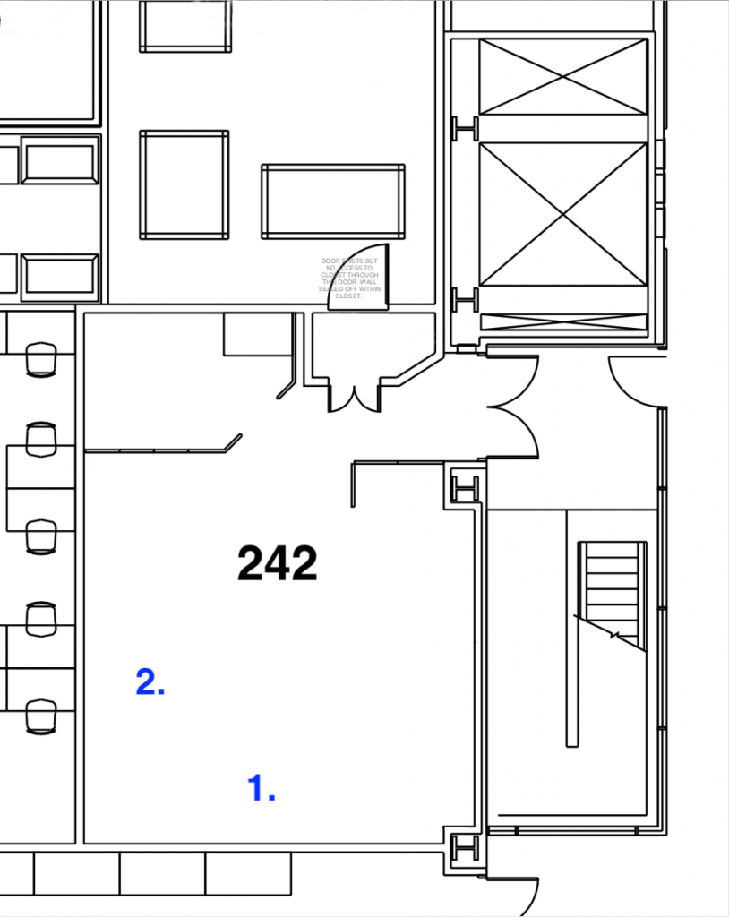
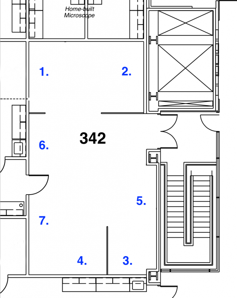
Third floor room 342
FAQ
Is network storage available?
Yes, CBS members are welcome to upload slide images to a web-based server (see link above). If logged in to the FAS Research Computing VPN server, microscope data can be saved to lab folders in \\hcbi5share.rc.fas.harvard.edu\share5\Other\CBS.
Is an image processing workstation available?
Yes, in the third floor imaging room.
Is there a tissue dissection suite?
Yes, through the door at the back of the third floor imaging room.
Is training required before using the microscope systems?
Formal training is required for the Nikon N-STORM. Ethan Garner’s lab manages training and scheduling for this system. Training is available for the other microscope systems but is not required.
What is the policy for the amount of time allowed on the microscope systems?
We have informal policy of up to 5 hours per day on a microscope system and up to 24 hours per week in total on all of the systems. Special arrangements can be made for imaging large tissue volumes overnight or for long-term time lapse imaging.