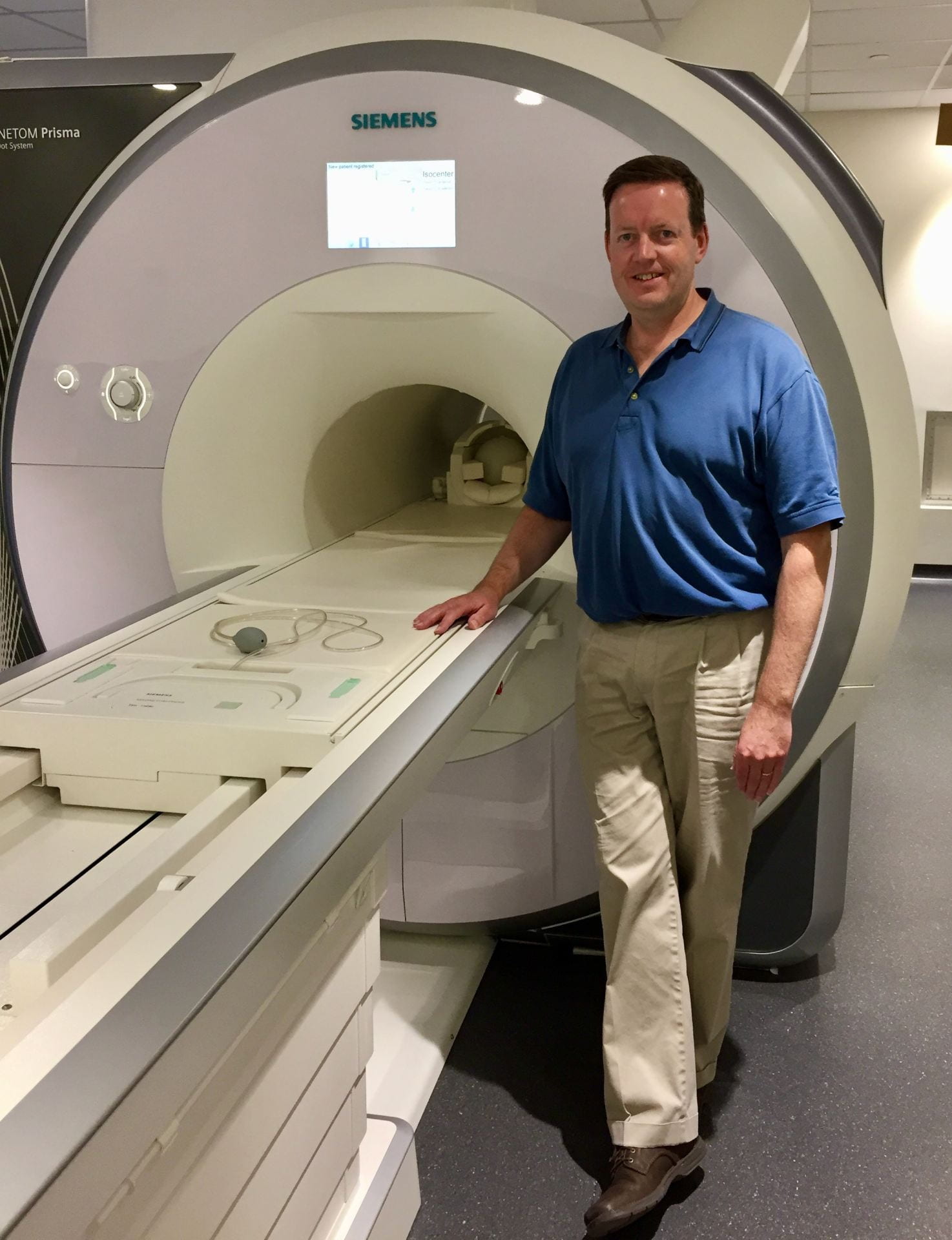
In August 2015, the CBS Neuroimaging Facility upgraded its 3.0 Tesla Siemens MAGNETOM Trio MRI scanner to the state-of-the-art 3.0 Tesla Siemens MAGNETOM Prisma whole-body MRI system (Siemens Healthineers, Erlangen, Germany). The Prisma MRI system has significant hardware advances and novel capabilities unavailable on other systems. This scanner has a short-bore (2m) magnet, a fast gradient system that provides high-speed structural and functional imaging, and a 64-receiver channel data acquisition system for parallel imaging with acceleration factors up to four-fold, permitting increased temporal resolution and reduced geometric distortions in functional MRI scans. The gradient rise time (200 T/m/s on all three gradient axes simultaneously) and peak gradient strength (80 mT/m per axis), and duty cycle are the most powerful specifications in the industry for whole-body systems (double the current standard gradient strength for advanced human MRI systems). Other novel features include a parallel transmit array to allow B1 shimming, zoomed image field-of-view selection, and selective excitation, all of which improve image uniformity and help to reduce image acquisition time; an all-digital RF chain between the control room and the magnet, which significantly improves RF stability and image SNR; enhanced parallel-receive array hardware, including a 64-channel head/neck array modified to improve subject comfort, increase their vision from the coil, and designed to permit simultaneous EEG or other external equipment, while increasing image SNR over the previous 32-channel coil design used on the former Siemens Trio MRI scanner; modern image reconstruction hardware optimized for rapid reconstruction of images requiring computationally intensive steps, allowing novel image acquisitions to display results in real-time rather than minutes/hours later or require manual offline reconstruction.
The Siemens 64-channel head/neck receive-array coil further enhances image SNR especially for functional MRI, with its significantly improved peripheral image SNR, generally corresponding to the cortex of the human brain. The 64-channel coil for the Prisma system has been designed to permit greater subject comfort with an increased visual field of view for the subjects, and greater accessibility for peripheral equipment including not only eye-tracking cameras, but for equipment that goes inside the coil, such as simultaneous EEG, transcranial direct current stimulation or transcranial magnetic stimulation. An improved 32-channel head receive-array coil and a 20-channel coil are also available.
The Siemens Prisma system is capable of EPI, second order shimming, CINE, MR angiography, diffusion and perfusion studies, and spectroscopy. The sequences available are matched to those of the MGH/HST Athinoula A. Martinos Center for Biomedical Imaging through a Master Research Agreement with Siemens.
Quality Assurance:
The performance of the Tim Trio system is monitored weekly for gradient linearity performance, and daily to ensure:
- minimal signal drift in EPI timeseries for fMRI,
- high temporal SNR in EPI timeseries for fMRI,
- consistent single image SNR for improved functional and structural MRI,
- minimal/consistent image ghost intensity on EPI and diffusion weighted images,
- lack of RF interference from other equipment that could cause image artifacts,
- lack of spiking due to gradient connections.
More details on the daily and weekly quality assurance scans can be found here.
In addition, we have begun using the fBIRN agar phantom on a 1-2 weekly basis to also document EPI time series stability, drift and temporal SNR in a manner consistent with other fBIRN sites. Results will appear soon.