Additional resources are available to investigators that can be used either in concert with MRI or for separate research studies
MRI Simulator (“Mock scanner”)
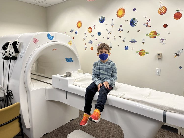
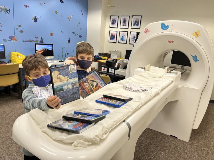
The MRI simulator or “mock scanner” suite is available to investigators to acclimate research subjects, prepare experimental paradigms, and train research staff for actual MRI studies. The mock magnet simulates the real MRI scanner and introduces research subjects, including normal and clinical populations, children and adults, to the scanning environment. This permits subjects to gradually become accustomed to the scanning procedure and trained to minimize movement. Allowing subjects to acclimate to the scanner before the actual imaging session helps to prepare them, and encourages calmer and more focused performance, which ultimately results in more productive and cost effective scanning sessions. The simulator can also be used to develop and test experimental paradigms, and to quickly train new researchers to run the experimental session in a safe and controlled environment, minimizing the use of costly scanner time for set-up and training.
Our mock magnet (NordicNeuroLab) has a bore diameter of 22” (56cm) (the height from the table to the top of bore is 16.5” (42 cm)), and utilizes a simulated head coil assembly with mounted mirror system. The simulator is equipped with a sliding patient table, realistic in-bore fans and lighting, and a sound system that mimics the vibrations and pulse sequence noises of an MR imager.
The experimental setup includes, headphones and rear-mounted monitor for auditory and visual stimulus presentation. Experimental stimuli are presented via laptop computer supplied by the research team. Computer generated stimuli are simultaneously presented to the subject and to an additional monitor and headphone set for the experimenter.
Two five-key button box response units, identical to those in the MRI scanner, enables researchers to better develop and test experimental paradigms in the mock scanner.
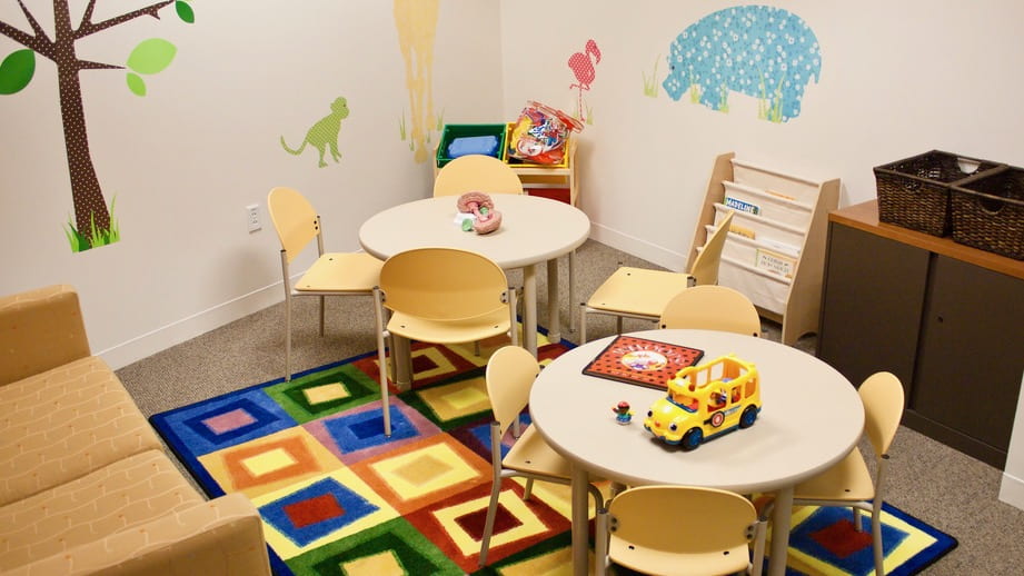
Child-friendly scanner decorations, and a head-set that detects movement and shuts off the video presentation are available for training young children. A comfortable observation area allows family members or caregivers to view the procedure from inside the mock scanner room.
MR-compatible Eye-tracker
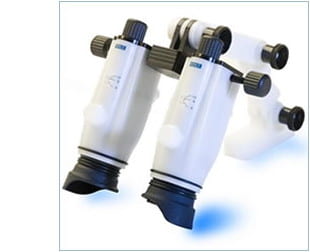
Our EyeLink 1000, from SR Research, is the most powerful eye tracker on the market, and has been customized specifically for the MRI environment.
The core of the EyeLink 1000 eye tracker consists of a custom designed high-speed camera connected to a dedicated host computer. Running on a real-time operating system, the host software provides extremely fast eye sample access with incredibly low inter-sample variability, accessed via a set of programming interfaces for multiple operating systems and programming languages.
We employ the Long Range Mount for the EyeLink 1000 with the Fiber Optic Camera upgrade that has been designed for MEG, MRI, and other applications that require the optics to be up to 150 cm from the eye, with low interference, and have a non-ferromagnetic optimized design. The Remote/Head Free camera upgrade option adds the ability to use the eye tracker as a fully remote system that does not require head stabilization.
The Fiber Optic camera uses the same internal sensor as the standard EyeLink 1000 camera, resulting in identical technical specifications as the standard camera for a given mounting option. When used with the Long Range mount, the Fiber Optic camera provides a non-ferromagnetic optimized solution for the MRI environment with a sampling rate and resolution that is unparalleled.
We hold periodic tutorial sessions where the set-up and use of the EyeLink 1000 system are demonstrated to users with interest in adding it to their studies.
Key Specifications (in remote/head-free mode, as used in the MRI environment)
Sampling Rate: 500 Hz Monocular
Accuracy: 0.5º average accuracy
Resolution: 0.05º RMS, saccade resolution of 0.25
Real-time Data Access: 3 msec (SD < 1.2 msec) @ 500 Hz
Participant Setup: Very simple and easy. Typically less than 2-5 minutes.
More details, and full specifications on the EyeLink 1000 can be found here.
Visual Stimulation
All visual stimulation for subjects during fMRI experiments is provided from the researcher’s own laptop computer. The facility does not provide a computer for this purpose. However, we do employ two different visual stimulation delivery systems at the CBS Neuroimaging Facility.
The first, and most commonly used, is a standard video-projection technique. The video display from the user’s laptop is sent via VGA cable to a conventional video projector, positioned behind the scanner room. It projects the display into the scanner room through a large feed-through tube, where it is projected onto a white plastic screen that bolts into the bore of the magnet. Subjects see the screen via mirrors attached to the head coils while in the magnet. This video delivery method is completely compatible with the EyeLink Eyetracking system. The video feed from the laptop is split so the researchers can also view it on a second monitor in the control room at the same time the subject in the magnet views it. Additionally, this video signal can also be viewed on the large-screen in the control room for teaching purposes.
For more advanced video requirements, the NordicNeuroLab VisualSystem is available.
The VisualSystem allows the researcher to easily present high-quality graphics or text to the subject. It has separate channels for each eye, and using frame sequential presentation, true 3D and stereo stimuli can be displayed. The VisualSystem has been designed with an adjustable arm that allows for fast positioning into the preferred angle of view for each subject, and easy securing the Siemens 12-channel head coil. It will not, however, attach to the 32-channel head coil, due to the design of the 32-channel coil. All studies using the VisualSystem must be conducted with the 12-channel coil at this time.
Because the VisualSystem completely covers the eyes, rendering it incompatible with remote eyetrackers like our EyeLink system, an integrated EyeTracking Camera is included with the VisualSystem. The core piece is an MR-compatible camera, which, thanks to a built-in infra-red light source, also works in darkness. The obtained composite video signal can be used to simply monitor, or, with the help of eye tracking software, to record and analyze eye movements. Our set-up provides a single camera solution for the monitoring of either the left or the right eye. NNL can provide, on request, binocular setups capturing the movements of both eyes.
Auditory Stimulation
With easy and accessible controls, the Siemens communication console allows two-way simultaneous patient communication and full flexibility of audio settings. As an alternative to the built-in microphone and speaker, the technician can choose between different headsets according to individual preferences. The console also allows for recording of oral responses from the patient using a fiber-optic patient microphone.
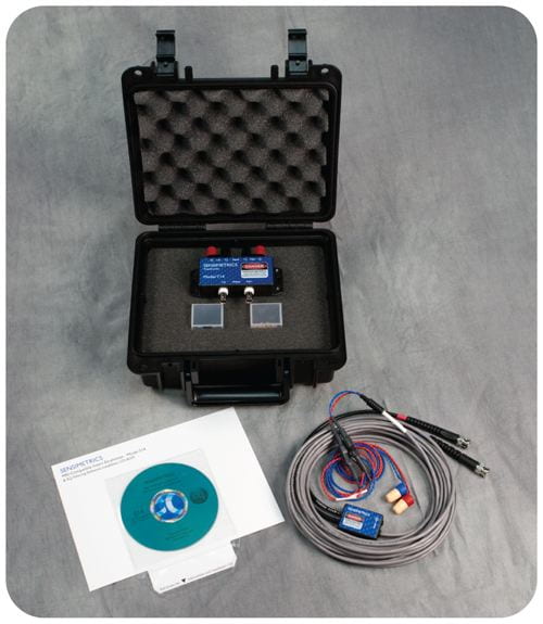
The S14 fMRI-Compatible Insert Earphones from Sensimetrics Corporation are designed to provide both high-quality acoustic stimulation and hearing protection from scanner noise. They are small enough to fit within any headcoil, and can be covered with circumaural muffs for added protection if the coil allows.
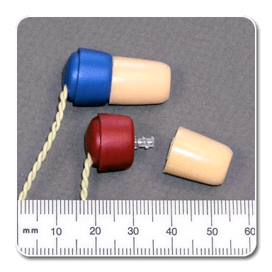
The S14 system takes audio (especially .wav files), and uses a third-party audio amplifier for amplification before transmission to the subject in the scanner. The transformer/ earphone combination will require no more than 3 watts per channel, which is met by a large number of small audio amplifiers. We use the Pyle Home PCA1 amplifier for this purpose. To ensure accurate frequency reproduction, we recommend use of the custom EQ Filtering software from Sensimetrics to modify your .wav files before playing them to a subject. This software runs on any Windows PC, and will ensure accurate frequency reproduction for our particular set of earphones. The EQ Filtering software is available from center staff./image
Physiological Monitoring
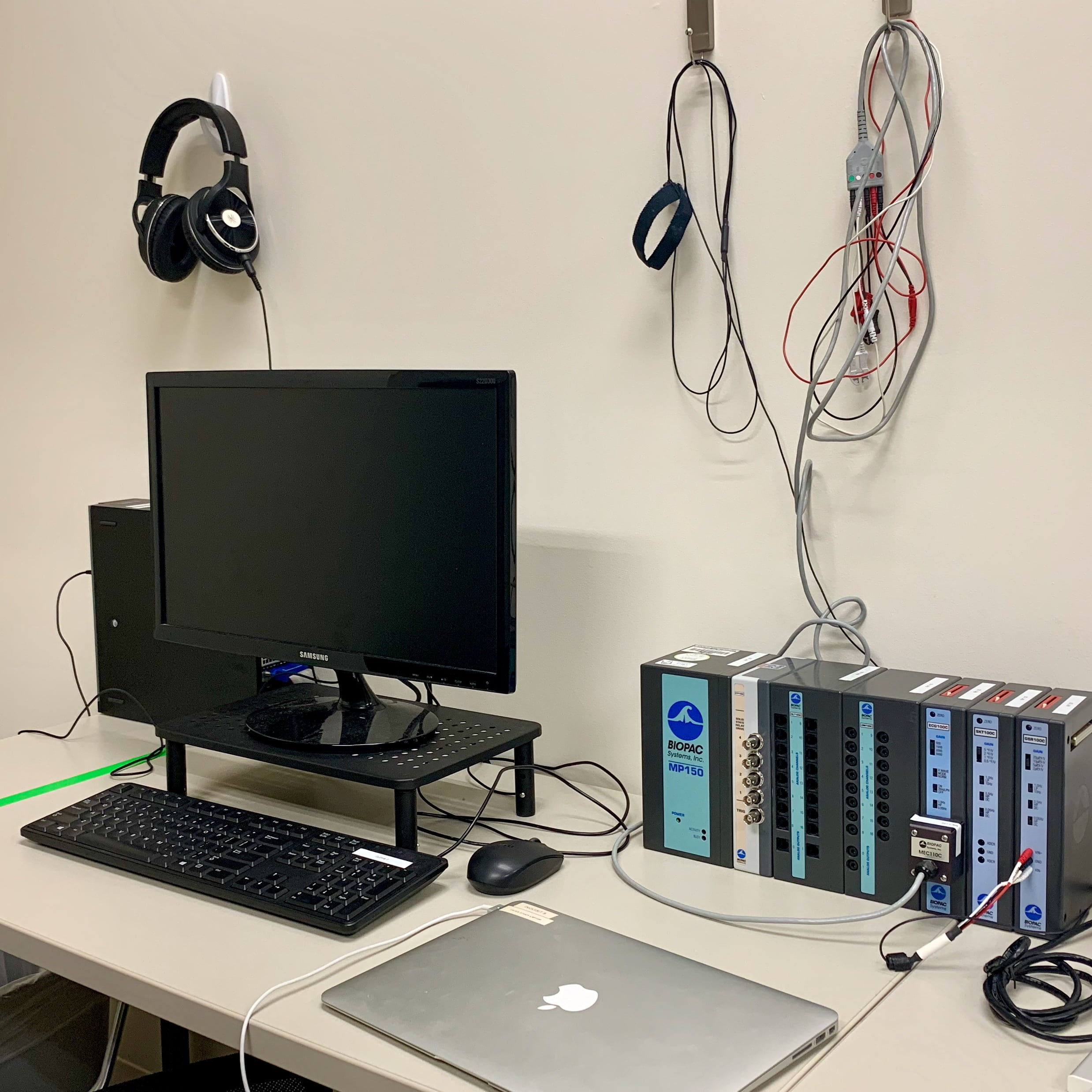
An MR-compatible BIOPAC MP150 system is available for MRI studies while a second MP150 system is located in the physiological testing suite. These modular data acquisition & analysis systems can be used for cardio-pulmonary and neuro-physiology measurements.