Image credit: Jeff Lichtman
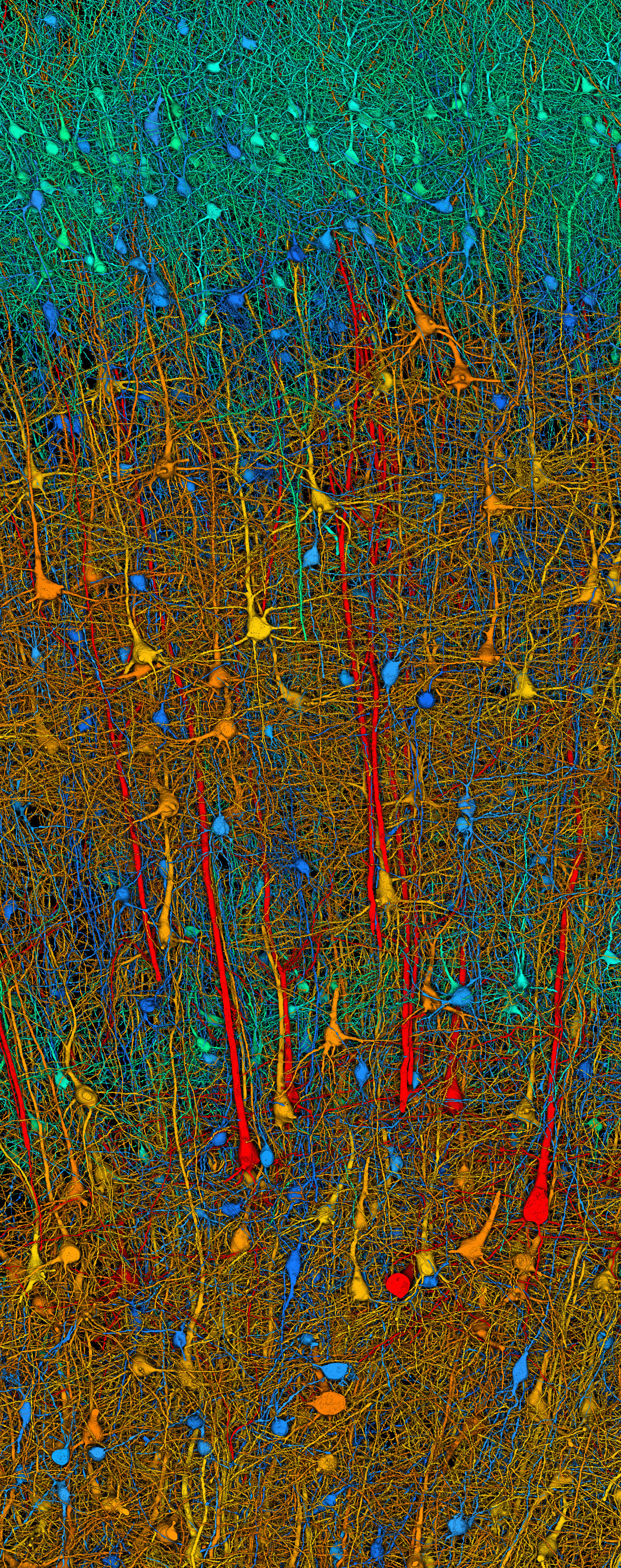
You can click on this image, which is the background image of the homepage, created by Daniel R. Berger in Jeff Lichtman’s group from serial section electron microscopy data. The data come from a sample of anterior temporal cortex of a living human patient. You can zoom in on the image with another mouse click, pan to see details, and zoom in or out further using (Windows: CTRL+ or CTRL-, MacOS: CMD+ or CMD-) on your keyboard.
Selected excitatory and inhibitory neurons are included here for visualization; the full data set includes all cells and parts of cells. The visible processes are overwhelmingly dendrites. Most of the processes belonging to neurons from another part of the brain have been removed for viewing clarity. The blue cells are large inhibitory neurons and their dendrites lack spines. The other colors code for the volumes of the excitatory neuron cell bodies: red > orange = yellow > green. Some axons can be seen as they leave the larger, excitatory neurons.
From top to bottom, the sample is 2.5 mm tall, 1 mm wide, and 165 microns thick. It runs from layer 2 of cortex down to layer 6.
The report of these results can be found here in Shapson-Coe et al.:
https://www.biorxiv.org/content/10.1101/2021.05.29.446289v4
The full data can be explored and studied in detail here:
https://h01-release.storage.googleapis.com/landing.html
https://h01-release.storage.googleapis.com/explore.html
https://h01-release.storage.googleapis.com/gallery.html